ICG Lymphography overview
Indocyanine green (ICG) lymphography is a specialised imaging test for lymphoedema. We inject a small amount of fluorescent dye (ICG) just under the skin and use a near-infrared camera to watch how the dye travels through your superficial lymphatic vessels in real time.
ICG lymphography helps confirm and stage lymphoedema and guides decisions about conservative treatment and surgery, including lymphaticovenous anastomosis (LVA), vascularised lymph node transfer (VLNT), and liposuction or selective debulking. It does not treat lymphoedema, but it provides detailed information that supports a tailored, physiology-led plan.
At a Glance
- Aim: Map superficial lymphatic vessels and flow patterns to help diagnose and plan treatment for lymphoedema.
- Best suited to: Suspected or confirmed lymphoedema where we need detailed staging or surgical planning.
- Setting: Outpatient procedure in a clinic or day-case setting.
- Anaesthesia: Usually none; sometimes a small amount of local anaesthetic.
- Duration: Typically 45 to 90 minutes, depending on the areas we image.
- Scars: No cuts; only tiny injection marks that fade over several weeks.
- Safety: Very small dye dose; serious reactions are uncommon. Previous severe reactions to ICG, iodine or shellfish allergy need careful discussion.
About ICG lymphography
01How ICG lymphography works
ICG lymphography uses a fluorescent dye and a special camera to show how lymph flows through the superficial lymphatic system.
In practical terms:
- We inject a small amount of ICG just under the skin at specific points, usually near the hand or foot and sometimes at additional sites.
- The dye enters nearby lymphatic vessels.
- A near-infrared camera detects the dye as it moves, so we see bright lines where lymphatic vessels carry the dye and different patterns where flow slows, leaks, or stops.
- We acquire images over several phases to understand both the pathways and the speed of lymph transport.
The test shows:
- Where lymphatic vessels remain open and carry fluid forwards.
- Where lymph starts to backflow into the skin (dermal backflow patterns).
- Areas with very limited or absent superficial lymphatic flow.
This information shows how your limb drains and which parts of the limb show early, mixed, or advanced lymphatic damage.
02What does ICG lymphography add to your care?
ICG lymphography links what we find in the clinic with your treatment plan. It refines decisions that we already base on your history, examination, and previous tests.
ICG lymphography helps us to:
- Confirm and stage lymphoedema and distinguish it from other causes of swelling.
- Decide whether options such as LVA, VLNT, liposuction or debulking are appropriate, or whether conservative care alone remains best.
- Monitor change after surgery or during conservative care and, in selected people after cancer treatment, support a surveillance plan.”
ICG lymphography supports a physiology-led, tailored approach rather than a one-size-fits-all pathway. In practice, the value of ICG lymphography lies in how we interpret the patterns and how they change your plan. At the Great North Lymphatic Centre, we interpret early and later phases of your scan together and use this information, alongside your symptoms, examination, and goals, to decide whether options such as LVA, VLNT, liposuction, or conservative care are likely to help.
03Who might benefit from ICG lymphography?
ICG lymphography may help if:
- You have swelling that we suspect represents lymphoedema, and we need to confirm the diagnosis and stage.
- You have known lymphoedema and want to explore surgical treatment options such as LVA, VLNT, or lymphoedema liposuction and debulking.
- You have mixed or complex lymphoedema and we need to understand which regions still have usable lymphatic vessels for planning.
- You have had previous lymphoedema surgery, and we need to understand which areas still have usable lymphatic vessels for planning further procedures or assessing the current pattern.
You may not need ICG lymphography if:
- Your swelling clearly relates to another cause other than lymphoedema.
- You have very advanced lymphoedema where imaging would not change our advice or plan.
04Benefits and limits
Potential Benefits
ICG lymphography can:
- Give a clear visual map of how your limb drains, which many people find helpful in understanding their condition.
- Support a more confident diagnosis of lymphoedema when examination or previous tests have left some uncertainty.
- Show which regions still have functioning superficial lymphatic vessels and which are more severely damaged.
- Help us choose whether options such as LVA, VLNT, liposuction, debulking, or conservative care are likely to help, and how to sequence them.
Guide focused compression or bandaging strategies and provide a record that you and your wider lymphoedema team can revisit over time.
Limits
- Shows mainly superficial lymphatic vessels; it does not assess deep lymphatics or veins, so some people still need ultrasound, MRI, CT or venous studies.
- It captures your lymphatic function at one point in time; patterns can change as disease progresses or treatment alters.
- Image quality can vary with skin thickness, scarring or previous radiotherapy
- It does not treat lymphoedema; it informs diagnosis and planning, and its value comes from how we use the information in your overall pathway.
What to expect
01Before your ICG lymphography
ICG lymphography usually takes place as an outpatient visit, following your first face-to-face consultation.
- You can usually eat and drink as normal unless we advise otherwise.
- Please bring a list of medicines, allergies and any relevant clinic letters.
- Bring your current compression garments; we may ask you to remove them briefly for imaging.
02During the test
The exact process depends on which limb or area we assess, but it usually includes:
- Explanation and marking
We explain the procedure, answer questions and mark the planned injection sites. - Injections
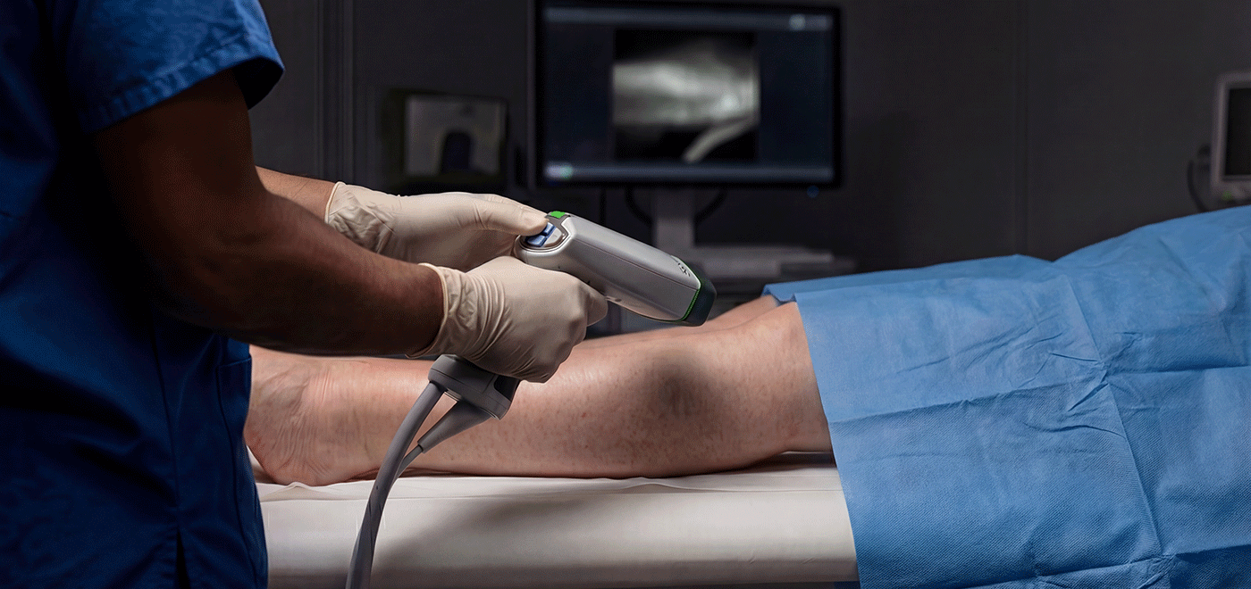
We inject a small amount of ICG dye under the skin at several points. You may feel brief stinging or pressure with each injection, which settles quickly. - Imaging
Using a near-infrared camera, we observe how the dye travels through the lymphatic vessels in a low-light room. You may be asked to move or position the limb to aid assessment. We take images or short video sequences and may mark key pathways on the skin to guide future planning.
The whole process usually takes between 45 and 90 minutes, depending on the areas we scan and how quickly the dye travels in your case. You would remain awake throughout and can ask questions during the test.
03After the test
- You can usually stand up, dress, and go home or continue your clinic visit soon after the test.
- You can usually drive and return to normal activities the same day.
- We review the images with you and explain what they mean for your diagnosis and treatment options.
04Risks and safety
ICG lymphography is generally well tolerated, but it carries some risks.
- Common, mild effects: brief stinging with injections and small bruises at injection sites.
- Temporary greenish staining: these marks usually fade over 2–3 weeks.
- Allergic reactions: serious allergic reactions to ICG are rare but can occur. We screen for previous reactions and relevant allergies and monitor you during the test.
Common questions
01Is the test painful?
Most people describe brief stinging or pressure with the small injections, followed by mild discomfort at the injection sites. The camera does not touch you and does not cause pain.
02Does ICG lymphography use radiation?
No. ICG lymphography uses near-infrared light rather than X-rays. It does not expose you to ionising radiation.
03Can I drive or work afterwards?
In most cases, yes. The test does not involve sedation or general anaesthesia. If we plan any other procedure on the same day that affects driving, we will tell you in advance.
04How long does the test take?
Please allow around 90 minutes for consent, injections and imaging. If ICG lymphography takes place on the same day as a full consultation, the visit may last up to two hours.
05Will I need to change my compression for the test?
We may ask you to remove compression shortly before imaging so we can see the limb clearly. You can usually put garments back on afterwards. We will tell you if we need any specific changes to your usual compression routine for the test.
06Does ICG lymphography replace other scans?
Not always. It adds detailed information about superficial lymphatic flow. We may still use other tests, such as ultrasound or lymphoscintigraphy, if they answer different questions.
Next steps
To discuss your suitability for lymphoedema surgery, contact our team. We will review your history, current treatment and goals, and advise on the most appropriate next steps at our luxury medical centre at The Beverley in Gateshead.
Complete our contact form to discuss your suitability for lymphoedema surgery.